We report a rare and life-threatening complication of spinal instrumentation infection that progressed to a secondary pseudoaneurysm of the abdominal aorta. The patient presented with a persistent infection following prosthesis removal, and imaging unexpectedly revealed a pseudoaneurysm requiring urgent endovascular intervention. This case underscores the importance of multidisciplinary collaboration and rapid diagnostic and therapeutic response in the management of complex vascular complications.
Case Presentation
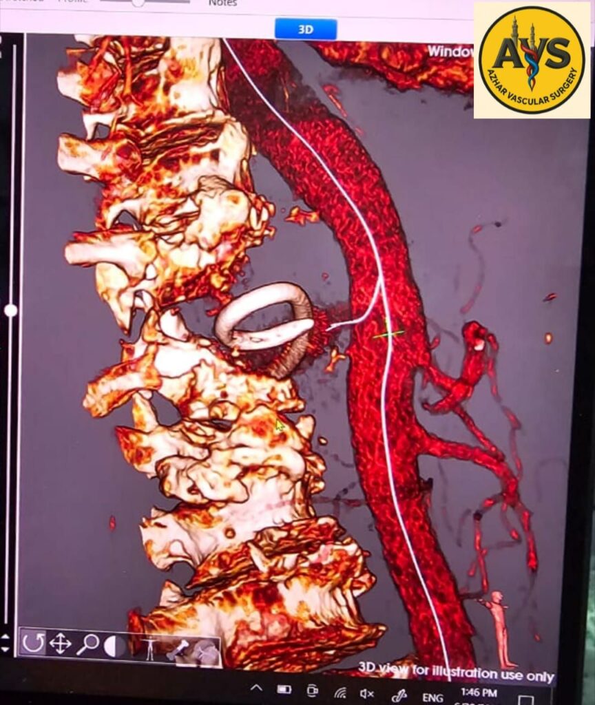
A 57-year-old male presented to Al-Sayed Galal University Hospital with a history of spinal fixation surgery using pedicle screws and rods, performed one year earlier following trauma to the back. Four months prior to presentation, the spinal prosthesis had been surgically removed due to postoperative infection. However, the infection persisted despite removal and conventional medical management.
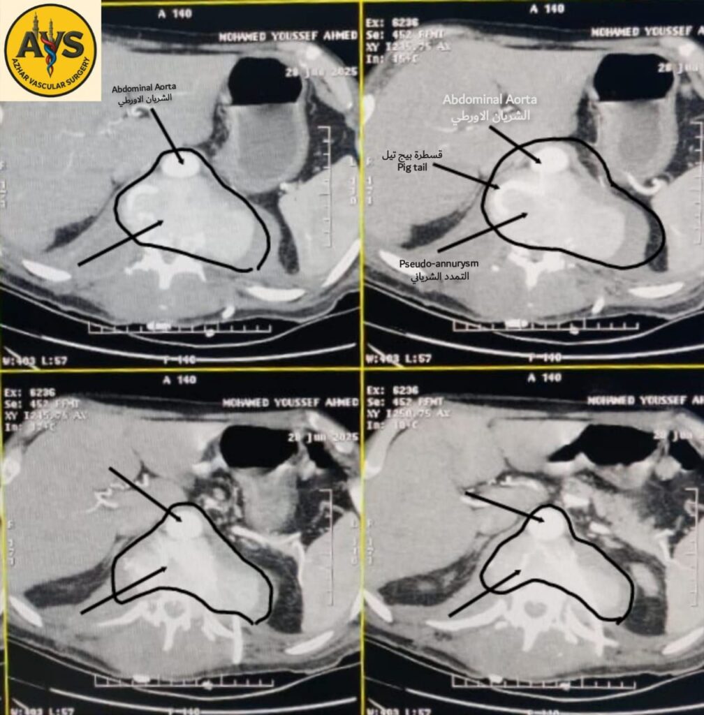
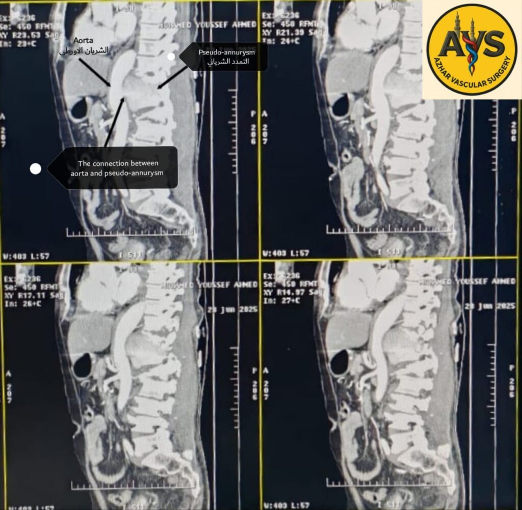
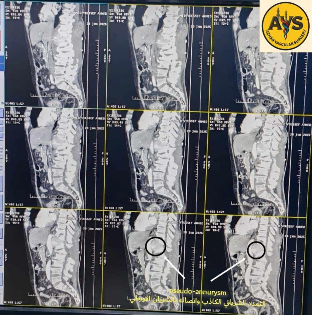
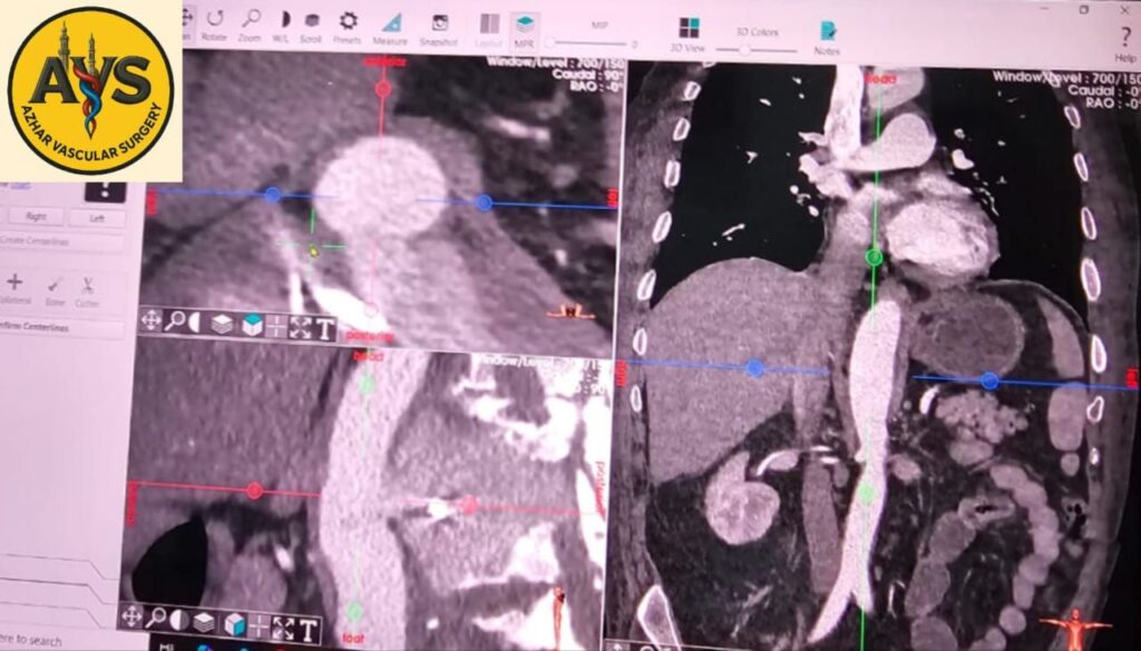
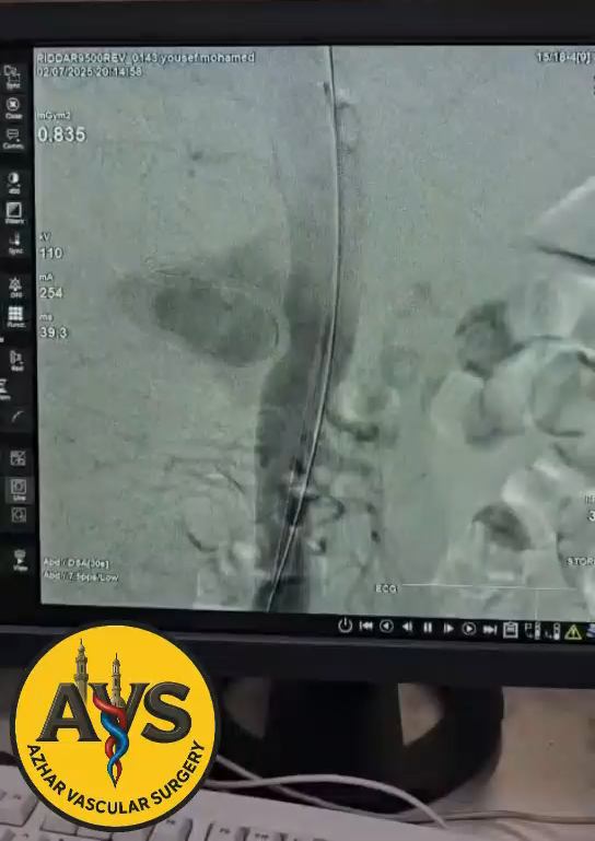
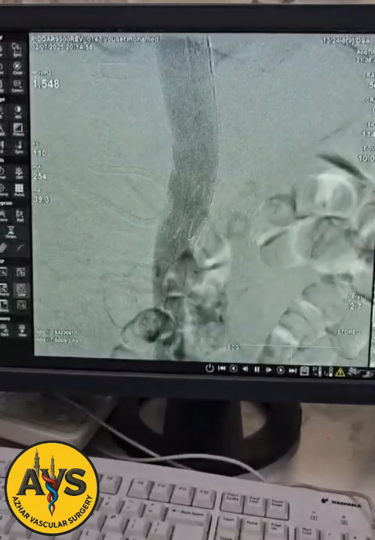
Magnetic resonance imaging (MRI) revealed a paravertebral collection displacing the aorta anteriorly, suggestive of an abscess. A pigtail catheter was inserted percutaneously under CT guidance to drain the presumed paravertebral abscess extending toward the para-aortic region. Unexpectedly, the catheter drained a large volume of arterial blood. Contrast-enhanced imaging subsequently revealed a pseudoaneurysm arising from the abdominal aorta.
Management and Outcome
The patient was immediately transferred to the operating theatre for emergency endovascular repair. A covered stent was successfully deployed to exclude the pseudoaneurysm, achieving complete hemostasis. The intervention was performed by a multidisciplinary vascular surgery team.
Conclusion
This case highlights a rare yet critical vascular complication secondary to spinal infection, emphasizing the importance of prompt vascular imaging in atypical clinical scenarios and the pivotal role of interdisciplinary teamwork in achieving optimal patient outcomes.
Written by : Dr/ Metawea.I.F